Spatiotemporal beam self-cleaning for high-resolution nonlinear fluorescence imaging with multimode fibres

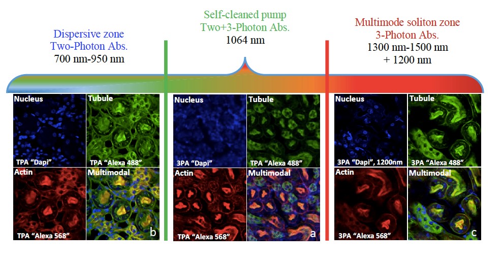
Beam self-cleaning (BSC) in graded-index (GRIN) multimode fibres (MMFs) has been recently reported by different research groups. Driven by the interplay between Kerr effect and beam self-imaging, BSC counteracts random mode coupling, and forces laser beams to recover a quasi-single mode profile at the output of GRIN fibres. Here we show that the associated self-induced spatiotemporal reshaping allows for improving the performances of nonlinear fluorescence microscopy and endoscopy using multimode optical fibres. We experimentally demonstrate that the beam brightness increase, induced by self-cleaning, enables two and three-photon imaging of biological samples with high spatial resolution. Temporal pulse shortening accompanying spatial beam clean-up enhances the output peak power, hence the efficiency
of nonlinear imaging. We also show that spatiotemporal supercontinuum generation is well-suited for large-band nonlinear fluorescence imaging in visible and infrared domains. We substantiated our findings by multiphoton fluorescence imaging in both microscopy and endoscopy configurations.
Reference:
N.O. Moussa, T. Mansuryan, C.H. Hage, M. Fabert, K. Krupa, A. Tonello, M. Ferraro, L. Leggio, M. Zitelli, F. Mangini, A. Niang, G. Millot, M. Papi, S. Wabnitz and V. Couderc, “Spatiotemporal beam self-cleaning for high-resolution nonlinear fluorescence imaging with multimode fibres,” https://arxiv.org/abs/2010.09340